The reproductive system refers to a system which consists of sexual organs responsible for reproduction and procreation. The parts of the organs of the reproductive system work together for procreation purposes, and they can either be internal or external. Owing to the vital role the reproductive system plays in procreation and continuity of the human race, it is considered by scientist as one of the most critical systems in the human body.
The Male and the Female Reproductive System
There is a big difference between the male and the female reproductive systems. All parts of the reproductive system in either men or women have distinct roles to play, and they can be grouped into primary and secondary organs. Primary organs are the testes and the ovaries because they are responsible for the production of sperms, ovum, and reproductive hormones. Other organs are grouped in the class of secondary organs because of their central role in facilitating the growth and maturation of gametes, delivery, and upkeep. It is also important to note that the parts of the reproductive system have different shape, structure and specific roles. All the reproductive parts in the female are located within the pelvis, but in males, some of them are outside the pelvis. In the next paragraphs there is a full discussion of the parts of the male and the female genital system.
The Female Reproductive Anatomy
The female reproductive system is designed to accomplish several reproductive roles in the production of eggs cells commonly known as the ova. The system is well designed to make it possible for the movement of the ova from the ovary to the fertilization site. Fertilization takes place at the fallopian tube or the oviduct, and after the egg is fertilized, it implants itself on the uterine walls. If fertilization and implantation don't take place, the female system has been designed to shed the uterine wall that has been formed, in a process known as menstruation. The reproductive cycles are maintained by sex hormones produced by the reproductive system.
The organs that make up the female reproductive system can be classified depending on where they are located, which means that they are either external or internal. The organs that form the female reproductive system include the mammary glands, breasts, the vulva, vagina, oviduct, uterus, and the ovaries.
1. External female reproductive structure
A woman's external reproductive parts have two principal roles: receiving sperms from the man during sex and the protection of the internal organs from infections. The external organs that make up the female reproductive system are discussed below.
-
Labia majora: Labia majora is a large lip-like structure outside the body whose function is to cover and offer protection to other external sex organs. Labia majora can be described as large lips with a fresh surface and can be compared to the scrotum in the male. The labia majora houses glands that secrete sweat and oil, and at the puberty stage, they get to be covered with hair.
-
Labia minora: Labia minora is small lip-like cutaneous foldings found on the inside of the vulva, and they are also known as the vaginal lips or the inner labia. The folding minimally extends to a width of inches surrounding the vagina and the urethra. The primary role of Labia Minora is to protect the clitoris, the urinary orifice, and the vaginal opening, and it is important to note that its size, color, and shape dramatically varies from one person to the other.
-
Clitoris: The clitoris is the part at which the two sides of the Labia Minora converge, and it is the point at which women pleasure comes from during foreplay and sex. It is located slightly above the vaginal opening and the urethra and is the most sensitive female sexual organ because it hosts thousands of sensory nerve endings which respond to stimulation during foreplay. The prepuce is a fold of skin that covers the clitoris and is homologous to the foreskin at the penis.
2. Internal parts of the female reproductive system
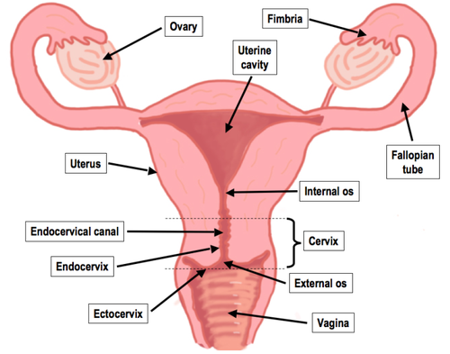
The principal internal organs in the female reproductive system are the vagina, the womb, the ovaries, and the fallopian tubes. The vagina and the uterus are semen receptors, while the ovaries produce ova and the fallopian tubes connect the ovaries to the uterus. Hormonal changes in the body cause one ova to be released from the ovary and moves down to the fallopian tube in a process known as ovulation. If the ovum is fertilized, implantation takes place in the uterus, and if not fertilized, the egg is eliminated through the menstrual process. The mentioned parts among others are discussed below:
-
The vagina: The vagina is a muscular tube that connects the uterus to the outside and acts as the penis receptor during intercourse. Through the vagina, sperms received from a man are carried to the uterus and then the fallopian tubes. The vagina serves as the passage for the baby during the delivery process, and for this reason, it is at times referred to as the birth canal and also acts as the conduit for menstrual flow.
-
The uterus: The uterus is referred to the womb in layman's language. It is a hollow muscular organ which is responsible for the development of the baby during pregnancy. The uterus is divided into two parts; cervix and the corpus where the former is the opening towards the vagina while the latter is the body of the uterus. The corpus can expand to the size of the developing baby, and it can contract to propel the baby outside the body during the delivery. The opening on the downward side of the uterus is known as the cervix, and it allows sperms to enter into the uterus and allows menstrual flow to exit.
-
The fallopian Tubes: Fallopian tubes refer to two muscular tubes which are attached to the upper section of the uterus where they serve as the passage for the ova from the ovaries to the uterus. The fallopian tubes are synonymous to the oviduct, and both the right and the left fallopian tubes have an ending in the shape of a funnel. The funnel-like structure is known as the infundibulum and is covered by finger-like projections called fimbriae. The fimbria picks up the released ova and then drop them to the infundibulum for delivery to the uterus. The sperm meets the ovum and fertilizes it in the fallopian tube after which it moves to the uterus for implantation in the walls of the uterus.
-
ovaries: Ovaries referred to a couple of small glands oval and located on the sides of the uterus. Ovaries play the role of producing eggs and sex hormones such as the estrogen and progesterone. Oocyte cells have the responsibility of producing ova which develop over time getting to maturity at puberty. During ovulation every month, a mature ovum is released which then travels to the oviduct for fertilization.
The Male Reproductive System
The male reproductive system is made up of sexual organs, glands and some duct systems that facilitate discharge of sperms. The functions of the male reproductive system organs are as listed below:
- Production, maintenance, and transit of sperms and semen
- Delivery of sperms to the female reproductive system during sex
- Secretion of male sex hormones to maintain the male reproductive system
1. The external organs of the male reproductive system
One remarkable difference between the male reproductive system and the female reproductive system is that most organs are located outside the body. Let's discuss the external organs of the reproductive system.
-
The penis: The penis lies outside the body and is used in sexual intercourse and consists of three parts. The penis reaches full size during puberty, and in addition to sexual functions, it serves the role of venting urine from the body. The three parts of the penis are the root, which is attached to the body at the abdomen, the shaft and a cone-shaped ending glans referred to as the glans. The glans has thousands of nerve endings and is usually covered by the foreskin, but the skin can be removed during circumcision. At the tip of the penis is the urethra which is a tube that helps in the transportation of urine and semen. The penis is made up of three chambers which consist of spaces that tend to fill with blood whenever a man is sexually aroused causing an erection. The erection of the penis makes penetration possible during sex, and the flow of urine is blocked to allow the flow of semen only. The tests are two organs that are in the shape of balls and exists on the outside of the body lying on the inside of the scrotum. The body of an ordinary man is supposed to have two tests which are responsible for the generation of sperm and the production of testosterone, which is the primary sexual hormone. On the inside of the testes are coiled tubes known as seminiferous tubules useful in the production of sperms. Testosterone hormone causes the urge to have sex and the development of bone and muscle mass.
-
The scrotum: The scrotum is a sac made of skin that hangs behind the penis and inside it lays the testicles, blood vessels, and nerve endings. The primary function of the scrotum is the regulation of temperature because normal development of sperm requires temperatures that are below body temperatures. The scrotum, therefore, acts as a climate control system aided by its muscles that easily contract and expand to move the testes closer or far from the body for optimum temperatures.
2. The internal organs of composing the male reproductive system
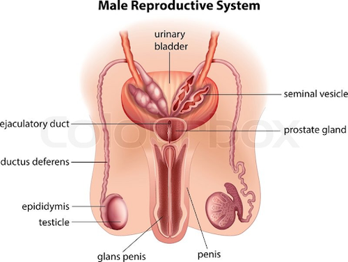
The parts that compose the internal organs of the male reproductive system are commonly referred to as accessory organs. Vas deferens, prostate glands, the urethra, epididymis, and the seminal vesicles are some of the primary internal organs as discussed below.
-
Epididymis: The epididymis is a duct or a tube that connects the testicles to the vas deferens and acts as the sperm storage area. It is made of long thin tubules tightly coiled to make a small mass that stores sperms till they mature, after which they can be delivered to other reproductive organs. The epididymis is long too and tightly coiled to delay sperms from being released to give them adequate time to mature.
-
Vas deferens: The vas deferens refer to a fibrous muscular tube that carries sperms from the epididymis and gives them a pathway to the urethra. The vas deferens has a broader diameter compared to the epididymis, and therefore the internal space helps in storage of mature sperms awaiting ejaculation.
-
The urethra: The urethra is a tube that gives passage to semen to the outside and passes urine from the urinary bladder. The urethra has a length of between 8 and 10 inches and passes through the prostate and ends at the tip of the penis. Urine and semen cannot pass through the urethra at the same time because the sphincters block urine from getting to the urethra during ejaculation.
-
Ejaculatory duct: The ejaculatory duct is the structure at which the vas deferens has joined the urethra. The ejaculatory duct opens during ejaculation to give sperms and secretions from seminal vesicles passage into the urethra.
-
Seminal vesicles: Seminal vesicles are sac-like pockets attached to the vas deferens at the base of the bladder. The leading role of the seminal vesicles is the production of fructose that provides sperms with the energy necessary for motion. The seminal vesicles fluid is the main component in the fluid ejaculated by man.
-
Prostate gland: The prostate gland refers to a gland located near the urinary bladder and the rectum. The gland produces a substance that is milky and alkaline which is useful in raising the ability of sperms to move and swim. The urethra passes through the prostate gland, and the components are emptied in the urethra to nourish the sperms.
-
Bulbourethral glands: Bulbourethral refers to small glands located on the inside of the urethra. The bulbourethral in response to sexual stimulation secrete an alkaline substance that neutralizes the acidity in the urethra due to residue urine and the vagina.
Fertilization in Human Beings
Fertilization refers to the joining of an ovum and the sperm to form a zygote which is the first stage in the development of a baby in the mother's womb. The union of the male and the female gametes takes place in the fallopian tube after which prenatal development starts. The process of fertilization is complicated as it involves various steps before resulting in real human life. The point at which fertilization takes place in the fallopian tube is known as the ampulla.
Fertilization is a process in which a sequence of events must be followed starting with the act of copulation where sperms are delivered to the female body. An egg is dropped from the ovaries and sits waiting for a sperm to fertilize it for life to begin. Therefore, the process of fertilization starts with ejaculation during sexual intercourse which is followed by ovulation and ends with fertilization. During copulation, the man releases millions of sperms which then race and the fastest sperm fuses with the egg after producing enzymes that make it possible to penetrate through the jelly coat of the egg. The sperm plasma fuses with the egg's plasma and then the head of the sperm disconnects after which the egg moves down along the fallopian tube until it reaches the uterus. Sperms produced and delivered to the female body should be able to complete the journey of meeting the egg in less than 48 hours after which they die.
Summary
The gender or the sex of a newborn baby is determined by the reproductive parts a child has after delivery. There is a big difference between the female reproductive system and that of the male reproductive system in with each of them playing distinct roles in reproduction. The organs of the reproductive system are divided into two; internal and external.